
3S is the abbreviation for “Seiwa Scholars Society,” which consists of the past and current Inamori Research Grant recipients. The 3S has evolved since 1997 with the hope that the interactions among the various specialties of the 3S members can lead to the further development of the research of their own. In the series “Visiting 3S Researchers,” we interview researchers in 3S who are very active in a variety of fields. Our ninth interviewee is Dr. Chikako Nagasato (2012 Inamori Research Grant Recipient) from the Muroran Marine Station, Hokkaido University.
Until now, we could not meet the 3S researchers in person because of the pandemic, but this time we were able to visit Dr. Nagasato at her laboratory, which was a first in this series of interviews.
Wakame (Undaria pinnatifida), Konbu (Saccharina japonica), Hijiki (Sargassum fusiforme), and Mozuku (a collective term for various types of brown algae from the family Chordariaceae)—These popular types of edible Japanese seaweed are all varieties of brown algae. They contain sticky components, which are often used for drugs and cosmetics. Growing to large sizes, brown algae play an essential role in maintaining the global environment and marine ecology. For this ninth interview with 3S researchers, we visited Dr. Chikako Nagasato, Director, Muroran Marine Station, Field Science Center for Northern Biosphere (FSC), Hokkaido University, to ask about her research on the reproduction and growth of brown algae.
Ecology of Seaweed Remains Wrapped in Mystery
──Could you tell us what led you to study brown algae?
Dr. Nagasato (title omitted below) I was originally interested in the fertilization process of animals and was enrolled at the Department of Biology, Faculty of Science, Yamagata University. When it came time for me to work in the lab, however, I realized that handling animals might not suit me. Given my lack of animal-handling aptitude, I could have worked on plants instead, but I didn’t have an advisor near me who was studying the fertilization of plants.
Wondering which path I should pursue, I happened to get an opportunity to join a training course offered by the Hokkaido University Muroran Marine Station (then the Institute of Algological Research belonging to the School of Science) in my junior year. During the course, I observed brown algae fertilization through a microscope, which piqued my curiosity as a researcher.
Matured brown algae release gametes into the sea water from the body, and these gametes can swim in the sea. Like animals, there are two types of brown algae gametes: female and male gametes, which correspond to animals’ ova and sperms, respectively. The former is fertilized by the latter. Female gametes adhere to rocks and such before male gametes do, and attract male gametes by releasing sex pheromones. Some types of brown algae have female and male gametes whose sizes differ greatly, like animals’ ovum and sperm, but other types have female and male gametes of similar size.
Having found brown algal fertilization so captivating, I decided to continue studying at the Hokkaido University Graduate School of Science to start my research work at the Muroran Marine Station, one of the few marine biological institutes in the world that exclusively deal with seaweed.
Generally, many species of seaweed, like brown algae, transform themselves over the course of a year. This is because of changes in generations, such as sporophytes and gametophytes in their life cycle, on top of seasonal changes. Konbu and Wakame, as we know them, are in the so-called sporophyte generation. Sporophytes disappear after releasing asexual reproductive cells, which eventually become gametophytes through successive cell division. Gametophytes of Konbu and Wakame are so small that they are almost invisible to the naked eye. Wakame and Konbu spend several months underwater as gametophytes, the former doing so until the sea gets cold in autumn and the latter doing so from autumn to early spring. If you go swimming in the summer at a beach along the main island in Japan, you don’t really see Wakame. This is because Wakame is in the gametophyte stage during that period.
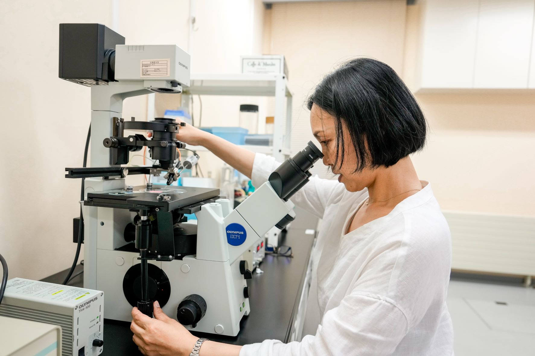
I’m particularly intrigued by tiny gametophytes and gametes before brown algae grow large. This is partly because of the ease of handling them in a laboratory, but I also enjoy imagining how such little things swim and get fertilized in the sea. I’m also curious about how a single cell from a fertilized egg divides itself to form a complex body.

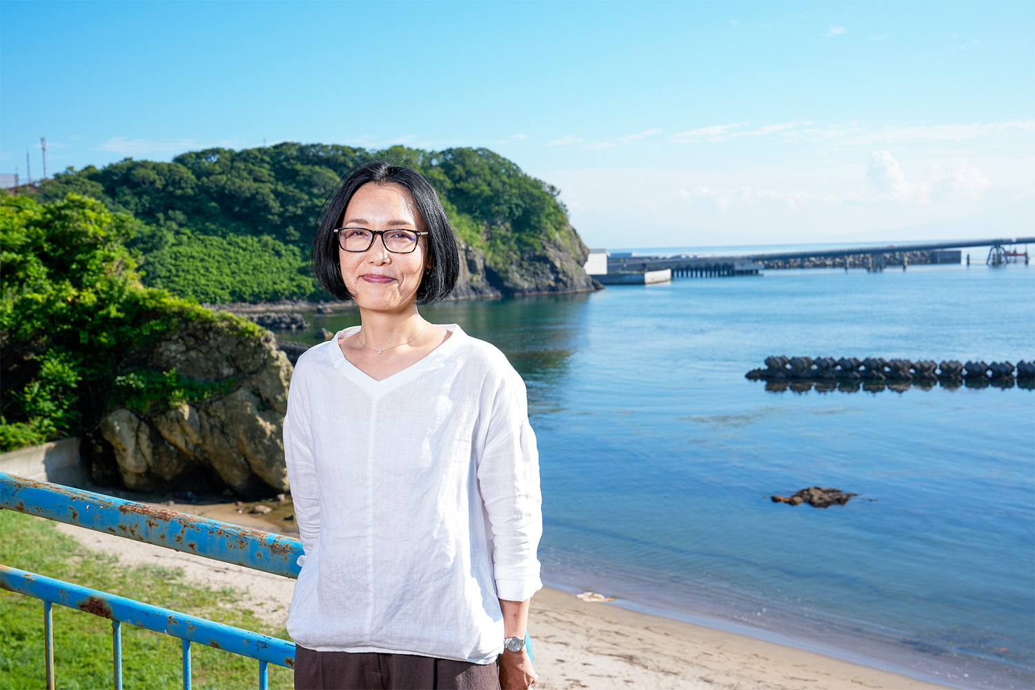
──Located near the ocean, the Muroran Marine Station is perfect for seaweed research.
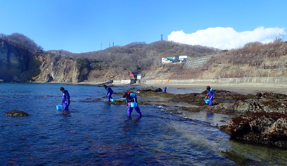
Nagasato We can always see the sea from the window of this room. Actually, a five-minute drive will take us to a place where we gather seaweed. We can also walk to the place. Some members use a bicycle, carrying buckets. Besides the proximity to our station, the place is ideal as we can gather a variety of brown algae there, with both cold and warm currents flowing into this coastal area to create rich seaweed vegetation.
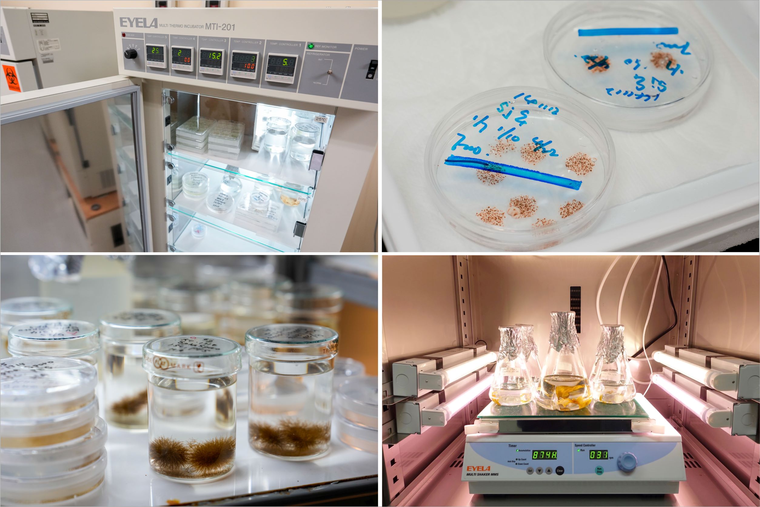
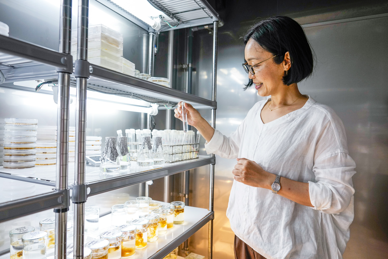
When studying seaweed’s life cycle, it’s more convenient to use laboratory cultures rather than gather seaweed that grows naturally. It’s really difficult to culture seaweed in a laboratory setting, but researchers here can take advantage of a wealth of diverse knowledge and experience that has been built up since the station’s establishment in 1933. We often host researchers from overseas who stay for a long period for research. We have an accommodation for them attached to the station.
──What has been the most exciting moment in your career as a researcher?
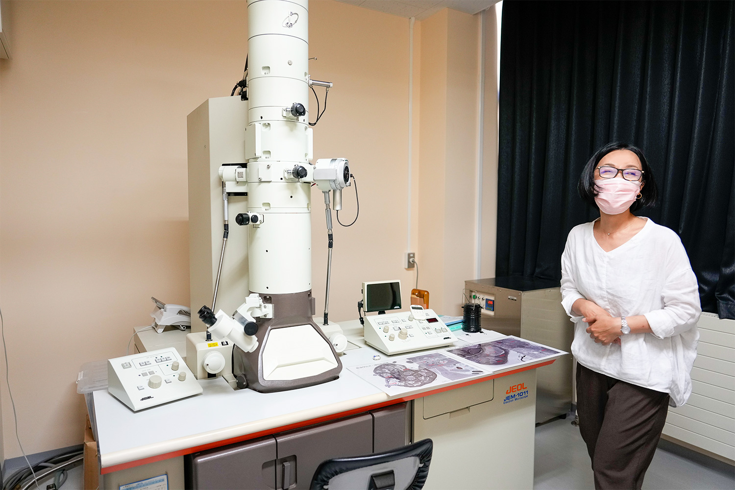
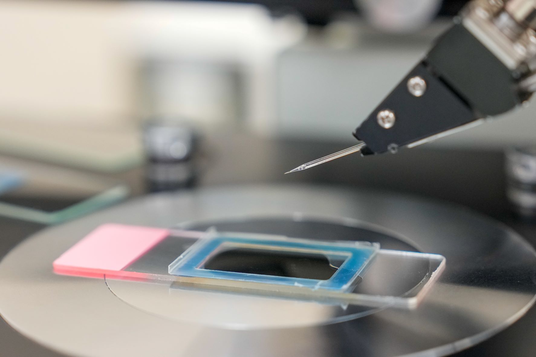
Nagasato It was when I captured a clear image of brown algal cell division by an electron microscope, which was different from the ones reported before.
Animals and plants follow different systems of cell division. Up to chromosomal duplication to divide them into two, the patterns are roughly the same, but they divide the cytoplasm differently. Animal cells are constricted in the middle and divided into two. Plant cells, on the other hand, aren’t; instead, something like a partition appears in the middle to divide a cell into two. When I started this research, it was believed that brown algal cells would divide after being constricted in the middle, much like animal cells. Many eukaryotes are divided into two after such constriction, and it was inferred that brown algae do the same. Furthermore, inside a brown algae cell is a cellular organelle called a centrosome, which can also be found in an animal cell. This cell organelle has a major role in the division of animal cells.
You would think that we could elucidate how cells are divided through clear images captured by an electron microscope, but it wasn’t the case because alginic acid, one of the components of brown algal cell wall, gets in the way of experimentation. Because alginic acid is viscid, fixatives or resins do not permeate samples when preparing sections for an electron microscope, which makes it difficult to gain high-resolution images. So, we did something to the cell fixation technique and reagents to develop a method to observe brown algae through an electron microscope, even with the presence of alginic acid. What we ended up finding was cell division similar to that of plants, with a division plate appearing in the middle.
──What does this discovery mean?
Nagasato Seaweed can be divided into brown algae, green algae, and red algae. Green algae and red algae, along with plants, are categorized into a group called the Archaeplastida, which are one of the major groups of eukaryotes. Brown algae, on the other hand, belong to a group called stramenopiles, which includes diatoms and oomycetes. Because many eukaryotes divide themselves by having their cells constricted of plasma membrane, this mode of division is supposed to be a prototype, but plants and brown algae might have acquired a similar but unique mode of cell division in their respective evolutionary process. I suspect the mode changed into one with the outgrowth of a cell plate through the process of wall-surrounded cells growing larger or becoming complex. With such a hypothesis bouncing around in my head, I work with my students to deepen our research.
How brown algae became a multicellular organism presents an interesting topic from an evolutionary perspective, too, because multicellular organisms need to ensure that their cells work in concert with each other. It is known that the cells of brown algae are connected to neighboring cells via a small tunnel-like structure, and we have confirmed that some substances are exchanged there. I predict that the nature of this exchange of substances via the tiny tunnels changes with shifts in the environment in which cells find themselves, as well as at different developmental stages. As we carry on this research, I’m also interested in how brown algae perform situation-dependent cell signaling.
Getting over the Difficulty Unique to Brown Algae Research
──Why do you think many things about brown algae remain unknown compared to plants and animals?
Nagasato It is partly attributable to the fact that not much knowledge has been amassed as there are few model organisms like mice and drosophila and that, compared to land plants, they are challenging to breed in artificial environments, which deters researchers from choosing seaweed as the subject for their study.
It’s only recently that their genome can be edited. In 2021, in joint research with our French collaborators, we were able to edit the genome of Ectocarpus siliculosus, which serves as a genomic model for brown algae, and presented a paper on this. We had long hoped to develop a tool to analyze how brown algal genes function by suppressing or increasing the expression of their target genes. To establish genome-editing technology, we have undertaken the process of trial and error for several years. Because brown algae cells have walls, however, it is more difficult to inject components needed for genome editing into their cells when compared with animal cells. Then at a conference held in France that I happened to attend, I knew that a group of French researchers was also working on this. As neither group seemed to be making progress, we decided to join forces and work together. It was only after we tried a microinjection device for plants that we were met with success.
Genome editing expands the range of what researchers can do. For example, we can use research findings for breeding, or capture cell behavior in real-time by manipulating genes of a cell structure that we wish to observe to make them fluorescent. Although we can only edit the genome of Ectocarpus siliculosus thus far, we are working to establish ways to do so for other types of brown algae too.
──If you were to pick a research collaborator, what background would you like them to have?
Nagasato I’m happy to collaborate with anyone, so long as they are interested in seaweed and have a specific idea for their research. My initial hope is to increase the population of seaweed researchers.
Asked what comes to mind when they think about seaweed, undergraduate students would reply by saying, “I like eating it” or “It’s gooey.” Apparently, it’s not really recognized as an object of research, and I hope to change that.
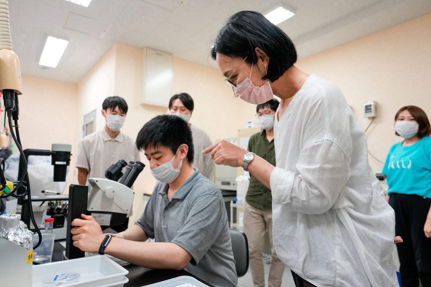
At the Muroran Marine Station, we organize a marine training course for students of the Department of Biological Sciences, School of Science, Hokkaido University, every May. They stay at the attached accommodation to conduct a series of tasks needed for seaweed research, such as experimentation of seaweed fertilization and growth, learning how to use fluorescence microscopes or electron microscopes, and gathering seaweed from the sea as a form of fieldwork. We also offer courses in spring and summer for undergraduates and graduate students from other universities in Japan or overseas. Some students participate in a summer course and return the following year to do the same course again.
Interest of Research on Seaweed You Can Work on with Various Styles
──Do you ever dive in the sea to gather seaweed?
Nagasato To tell you the truth, I can’t swim or dive. I also get seasick so I can’t sail out to the sea to do research on a survey boat. (laugh) Seriously, one of the researchers at the station is a qualified diver. Unfortunately, he can’t swim, either, but he can dive. After all, we are free to do research in any style we can.
──I must say it was interesting to learn that a seaweed researcher can’t swim!
Nagasato I understand how you feel. Born in Iwate Prefecture, I don’t feel comfortable unless I have easy access to the sea, but I can’t swim. I don’t like to eat fish so much, either, though I love eating seaweed as much as I love studying it, so I put seaweed into every dish I cook as much as possible. I also love going to places with ocean views. My favorite place is Etomo Cape, which is about a 15-minute drive from Muroran train station. I drive by the place on my way back home, and the setting sun over the sea is simply spectacular.
──Do you have a favorite seaweed?
Nagasato Yes, it’s a type of brown algae called Fucus. I have an emotional attachment to it, as the first fertilization of brown algae I saw was Fucus’s. This variety of brown algae only grows in cold places, and Muroran is its southernmost habitat, but I see them less and less in recent years, possibly because the southern limit is moving northward by inches. When I take my students with me to gather seaweed, I ask them not to gather Fucus. It’s ironic, but Fucus is now a rare species of seaweed though I came to Muroran to study it. Fortunately, it is still abundant in European seas
──Lastly, what message would you send to young students who aspire to be researchers?
Nagasato I want them to take an interest in a lot of things, not just their own research. I always tell students who are affiliated with this station to act proactively as, being away from the Sapporo Campus, they have few opportunities to communicate with researchers from other fields. I also tell them to contact professors from other laboratories directly by emailing them if they have something to ask them. It doesn’t matter if they need to contact Japanese researchers or otherwise.
Here at the Muroran Marine Station, the main areas of study are cell biology and the physiology of brown and green algae. I became involved in this field of research, initially fascinated by the fertilization of seaweed. I must say that seaweed has many aspects to explore, and it offers ever-lasting appeal. Seaweed is also important for our living and the global environment. I would love to see more people take an interest in research on seaweed.
| By My Side | Dr. Nagasato’s experimental tools On the tweezers are labels with her name on them. “I have used these tweezers since my first year at graduate school. Without these, I can’t hold tiny seaweed. The needles in the middle (third and fourth from left) are made for dentists, and I sharpen them with a nail file. To the far right is a very expensive instrument called Noyes scissors, which my advisor gave me when he retired.” 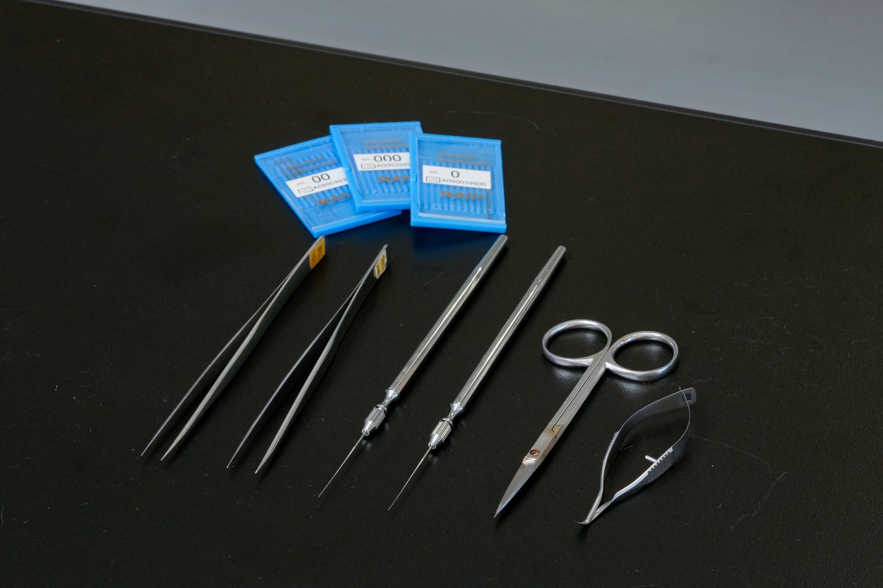 |
|---|---|
| This Book | Diversity and Evolution of Algae, compiled by Prof. Mitsuo Chihara, Shokabo Co., Ltd. Published in 1999, this is the textbookish reading for seaweed researchers. “This book gathers together almost everything we need to know about algae. Our previous Director, Taizo Motomura, contributed a column within. I assume that every algae researcher has a copy. Everyone at this laboratory has one. I invite students to a workshop to read this through.” 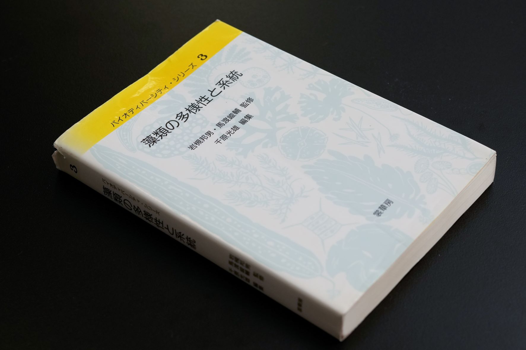 |
Chikako Nagasato
Professor/Director, Muroran Marine Station, Aquatic Research Station, Field Science Center for Northern Biosphere (FSC), Hokkaido University. After graduating from the Faculty of Science, Yamagata University, Dr. Nagasato matriculated at the Hokkaido University Graduate School of Science to study the reproduction and growth of brown algae at its Muroran Marine Station.
After becoming an Assistant Professor, FSC, Hokkaido University, in 2003, and an Associate Professor, FSC, in 2007, Dr. Nagasato took her current position in 2020.
Interview and original article: by Izumi Kanchiku (team Pascal)
Photo: by Kenta Nakamura