
3S is the abbreviation for “Seiwa Scholars Society,” which consists of the past and current Inamori Research Grant recipients. The 3S has evolved since 1997 with the hope that the interactions among the various specialties of the 3S members can lead to the further development of the research of their own. In the series “Visiting 3S Researchers,” we interview researchers in 3S who are very active in a variety of fields. The 18th interview is with Dr. Michihiro Suga (2016 Inamori Research Grants Recipient) from Okayama University.
Just as society relies on the activities of diverse people, in living organisms, countless proteins work together, each playing its own role to sustain life. Once we know which proteins work where and how, we will better understand the mechanisms of living organisms. The key to this understanding lies in the three-dimensional structure of proteins. Dr. Michihiro Suga of Okayama University has been analyzing the structure of microscopic proteins, partially revealing how photosynthesis works in the process. We visited his lab to learn the details of his research, which unveils the mechanisms of life through structural analysis.
How to Visualize the Structure of Minuscule Proteins
── How do you visualize the three-dimensional structure of proteins?
Dr. Suga (title omitted below) Proteins function like precision machinery within living organisms, and they are extremely small—approximately 2 to 10 nm in size. Given that human cells are approximately 0.01 mm in diameter, proteins are only about 1/2000 of their size. Because they are so small, they are not visible through regular optical microscopes. Since optical microscopes use visible light, with wavelengths of 400 to 700 nm, their resolution is not sufficient to resolve proteins, which are only a few nanometers in size.
That is why you need to use something with shorter wavelengths than visible light, such as X-rays or electron beams, to determine protein structure. X-rays have wavelengths of about 0.01 to 10 nm, which are similar to the size of atoms. This makes X-rays an excellent option for analyzing protein structures. However, the X-ray scattering signal from a single molecule or atom is extremely weak, so it cannot capture the structure on its own. Therefore, we crystallize the protein of interest so that its molecules are regularly arranged in the same orientation. By superimposing the scattering from a large number of molecules to amplify the signal, we can determine the three-dimensional structure of the protein.
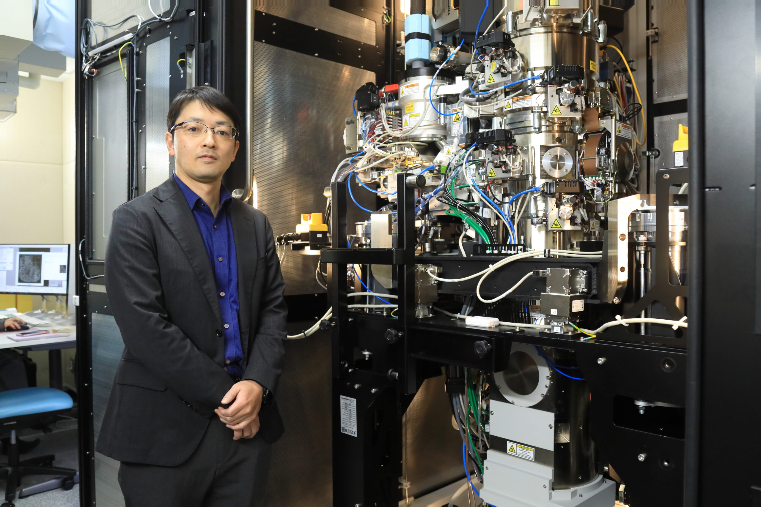
Suga Electron beams, on the other hand, have shorter wavelengths than X-rays. The limitations are that electron beams can only travel in a vacuum, and intense electron beams damage the proteins themselves. For this reason, conventional electron microscopy has relied on sample preparation involving dehydration, resin embedding, staining, and sectioning into thin slices. However, this approach made it difficult to observe proteins—which naturally function in solution—in their native state.
In recent years, a new method called “cryo-electron microscopy” has emerged, which allows us to analyze proteins in a near-native, aqueous state by rapidly freezing them. Thanks to this technological advancement, we can now unravel the structures of large proteins that are difficult to analyze using X-ray crystallography.
Cultivating Large Amounts of Algae for Experiments
── Preparing samples for observation using X-rays and electron beams must itself require a great deal of work.
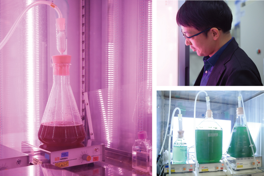
Suga That’s correct. The quality of the crystal directly affects the quality of the research. For example, when studying proteins involved in photosynthesis, we cultivate cyanobacteria (blue-green algae), a group of photosynthetic bacteria. From these bacteria, we extract the target protein and crystallize it. In research published in 2017*1, we cultured an amount equivalent to roughly 50 household bathtubs. Of course, we cannot culture them in bathtubs, so we cultured them in flasks, extracted the protein, purified it, and cryopreserved it—diligently repeating this process over and over.
── I had no idea you needed that much.
Suga In most cases, you may not. My research drew significant attention in 2015 when it was featured in Nature for elucidating the mechanism of the protein*2 essential for photosynthesis by analyzing approximately 1,000 crystals using an X-ray free-electron laser. We thought, if one crystal was not enough for analysis, why not use 1,000? Research published in 2024*3 also involved observing protein structural changes occurring within nanoseconds, which required preparing a massive amount of samples.
Luckily for us, many people say, “Your research is amazing,” but few would try to follow our approach. They might want to think of a smarter way (laughs). However, my philosophy is that as long as the research is logically sound and the experimental principles and design are reasonable, it is worth pursuing even if it requires considerable effort.
Elucidating Photosynthesis Through Structure
── What kinds of protein structures have you been studying?
Suga Among the various research projects I carry out, for many years I have been particularly focused on PSII, a protein complex essential to the water-splitting reaction in photosynthesis that generates oxygen from water. As for photosynthesis, we learned in elementary school that plants generate oxygen and nutrients from water, light, and carbon dioxide. On an atomic level, however, much remains unknown—in particular, how oxygen can be extracted from the stable water molecules has long been a subject of debate.
To explain this, we have been gradually revealing how PSII reacts and functions by elucidating its three-dimensional structure. First, we clarified the overall structure of PSII. Then, among the four intermediates through which its structure changes in response to light, we uncovered the structure of the final state immediately before oxygen generation*1. In 2024, we succeeded in capturing the transient states during the transitions between these intermediates*3.
── How significant do you think an understanding of photosynthesis is?
Suga In the long history of life on Earth, cyanobacteria are the only organisms that evolved to perform the photosynthetic process that splits water. Other living organisms, including plants and algae, evolved to perform photosynthesis by incorporating cyanobacteria into their cells. We believe that understanding how photosynthesis works helps us approach the profound mystery of how life emerged and, by applying these insights, brings us closer to achieving artificial photosynthesis.

── What do you find most fascinating about your research field?
Suga Uncovering protein structures often leads to unexpected discoveries and enables us to explain biological phenomena with precision. Even when we are unsure which of several hypotheses is correct, we can demonstrate a form of evidence by analyzing the structure. The structures we uncover often amaze me—some are simply beautiful, while others surprise me with their remarkable sophistication.
In recent years, AI technologies for predicting protein structures based on amino acid sequences have been developing rapidly. This may make you wonder if structural biology is no longer necessary, but that is not the case. AI is being trained on the vast amount of structural data that researchers have persistently accumulated through experiments. To improve the accuracy of these predictions, it is essential that we continue determining actual structures through experiments.
Current AI cannot predict the phenomena where proteins undergo transient fluctuations and become unstable, such as the intermediate steps of PSII reported by our group in 2024. This reminds us that humans still have an important role to play. At the same time, AI often provides unique insights. I believe that combining the strengths of AI and humans will further expand the possibilities of our research.
── What kinds of research projects do you want to pursue going forward?
Suga I hope to investigate more deeply into how photosynthesis works and apply the findings to help achieve artificial photosynthesis. Since Japan has limited natural resources, it would be wonderful if we could generate energy using sunlight and water.
That said, artificial photosynthesis is not something I can achieve alone. It will require cooperation from researchers across various fields, including chemical catalyst development and industrial engineering. I believe such interdisciplinary efforts are crucial and would like to continue engaging in them.
Aside from these efforts, I would also like to create something new within the realm of biology. For example, I am currently analyzing the structure of transporter proteins, which mediate the uptake of external nutrients into plants. I hope to apply the results to develop living organisms with new characteristics, such as crops that absorb fewer harmful substances and plants that can grow even in harsher conditions.
── Have you always wanted to become a researcher since you were a child?
Suga Not at all. As an elementary school student, I liked science, but when asked what subject I liked best, I would answer, “lunch break.” During my university years, I was in the judo club and devoted most of my time to practice; Honestly, I rarely attended classes. Once I joined the research lab, however, I threw myself into my research. Since I was in the Faculty of Science, few of my classmates were job hunting, and I simply followed the flow, which has taken me to where I am today. As a researcher, I can devote myself to what I truly enjoy without anyone stopping me, so I suppose it suits me well as a career.
── You advise the Judo Club at Okayama University. Do you still practice judo?
Suga Not anymore. But I started running to stay healthy and lose weight about a year ago. I run at the Okayama Prefectural Multipurpose Grounds, which is just a few blocks away from the campus. Running on the same trails in the same park, I can sense the seasons changing. In the spring, the cherry blossoms bloom, and I see lotus flowers just before the summer starts. In the autumn, I see the fall leaves along with ripe ears of rice. It is my favorite place because I can experience the seasonal shifts by observing the scenery from the same place throughout the four seasons.
*1. M. Suga et al. (2017) Light-induced structural changes and the site of O=O bond formation in PSII caught by XFEL. Nature 543: 131–135
*2. M. Suga et al. (2015) Native structure of photosystem II at 1.95 Å resolution viewed by femtosecond X-ray pulses. Nature 517: 99–103
*3. H. Li et al. (2024) Oxygen-evolving photosystem II structures during S1-S2-S3 transitions. Nature 626: 670–677
| By My Side | 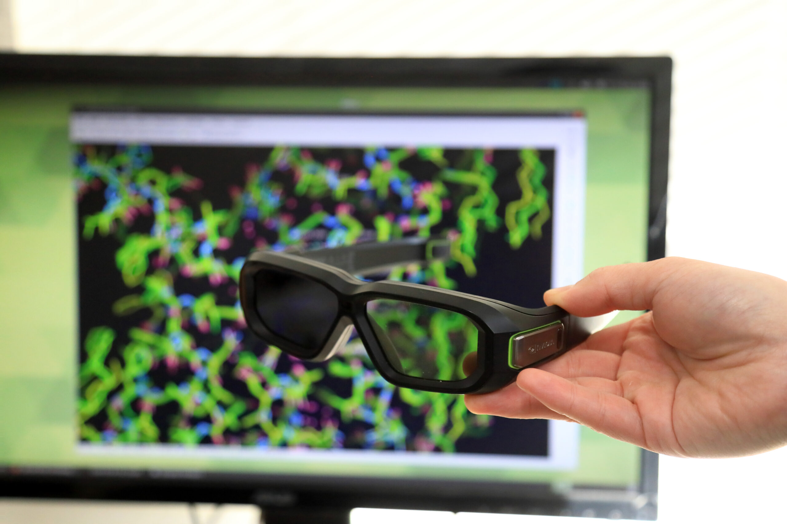
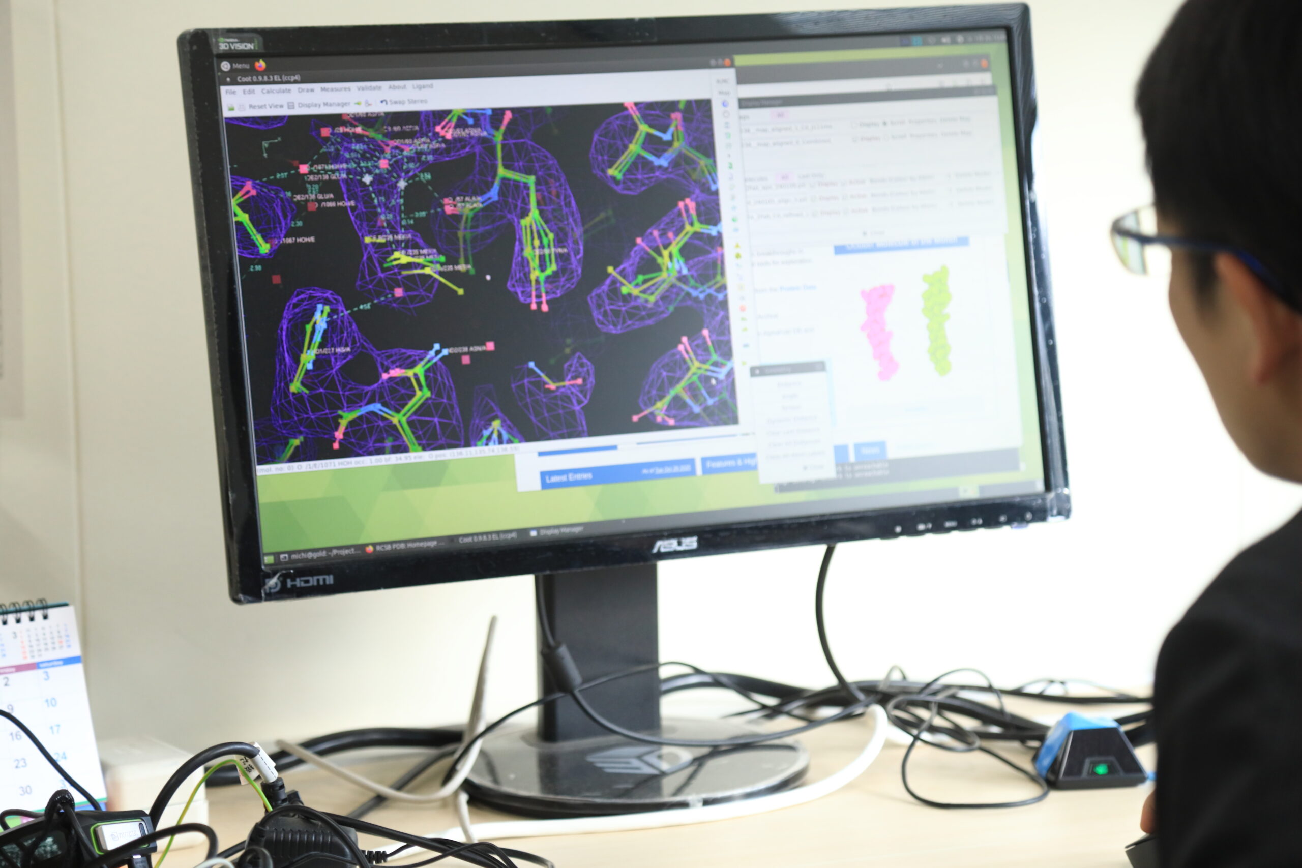
3D glasses 3D glasses help clarify the depth and contrast of the three-dimensional structures of proteins that are visualized using analysis results. “I observe images for quite a long time wearing these glasses. Sometimes, while I’m rotating the image of a protein’s three-dimensional structure, ideas suddenly come to me about how the protein functions and works.” |
|---|---|
| This Book | 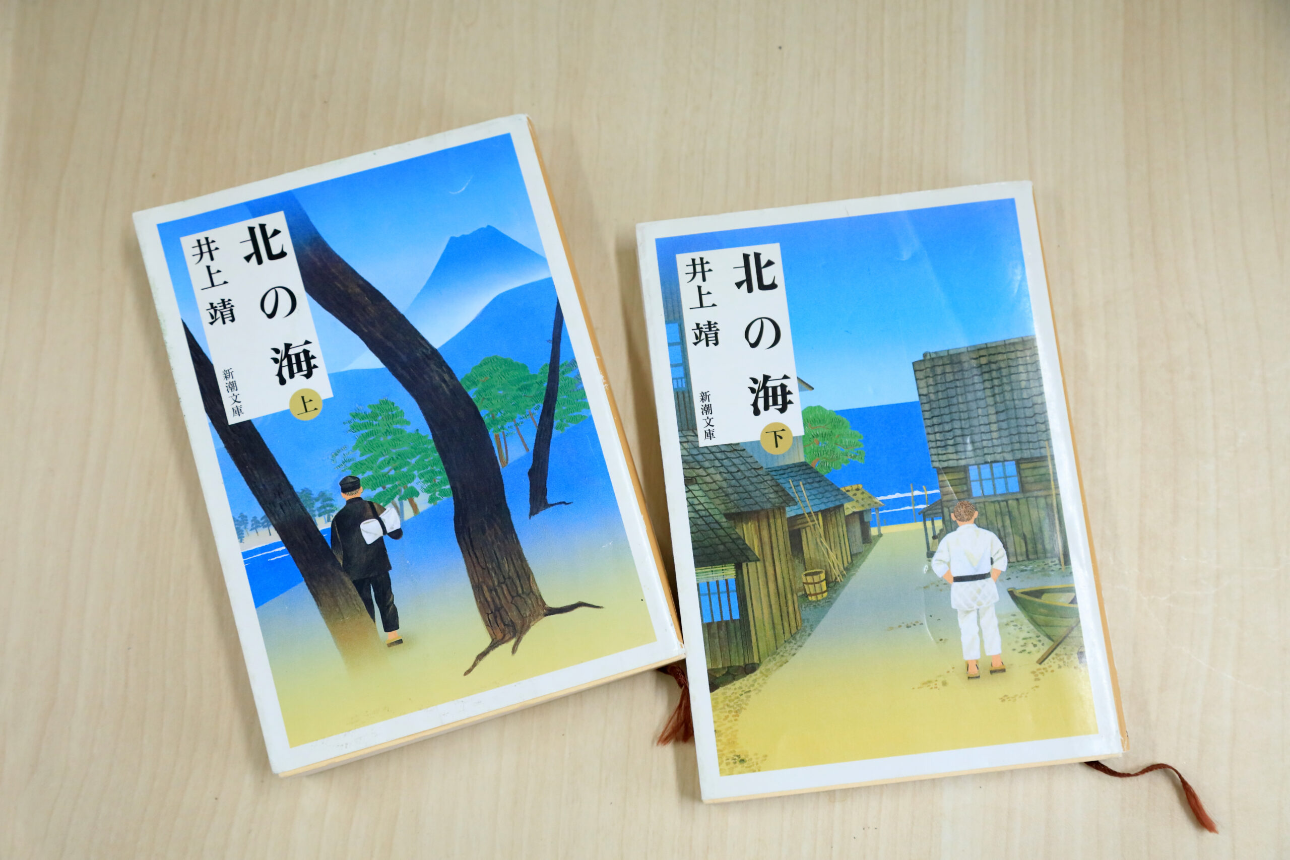
The Northern Sea (‘Kita no Umi’) by Yasushi Inoue, SHINCHOSHA Publishing Co., Ltd. (Book title translated) In college, Dr. Suga devoted himself to Kosen judo, a style focused on ground fighting. “I believe anyone practicing Kosen judo in the Shichitei (Seven Former Imperial Universities) judo clubs has read this book. The main characters’ rivals are members of the judo club of Dai-Roku Koto Gakko, the predecessor of Okayama University, so as someone from Okayama, I found this book relatable.” |
Michihiro Suga
Professor at the Research Institute for Interdisciplinary Science, Okayama University. Born in Okayama Prefecture. After completing his Ph.D. in Science from the Graduate School of Science, the University of Osaka, in 2009, he served as a postdoctoral fellow at the Institute for Protein Research, the University of Osaka, from 2009, and at Oregon Health & Science University in the U.S. from 2010. He then joined Okayama University as a Specially Appointed Assistant Professor in 2012, became an Assistant Professor at the Graduate School of Natural Science and Technology in 2014, and was appointed Associate Professor at the Research Institute for Interdisciplinary Science in 2016. He has been in his current position since 2022.
Interview and original article: Izumi Kanchiku (team Pascal)
Photo: Shina Matsumura
-
 Research Grants
Visiting 3S Researchers #18 Dr. Michihiro SugaCapturing Transient Conformational Changes in Proteins to Unravel the Mysteries of Life —Unveiling the Unseen Intermediate States of Photosynthetic Reaction—
-
 Research Grants
Visiting 3S Researchers #16 Dr. Taisei ShidaRestoring the Thoughts of Ancient Indian Philosophers from Texts —Identifying Errors in Palm Leaf Manuscripts—
-
 Research Grants
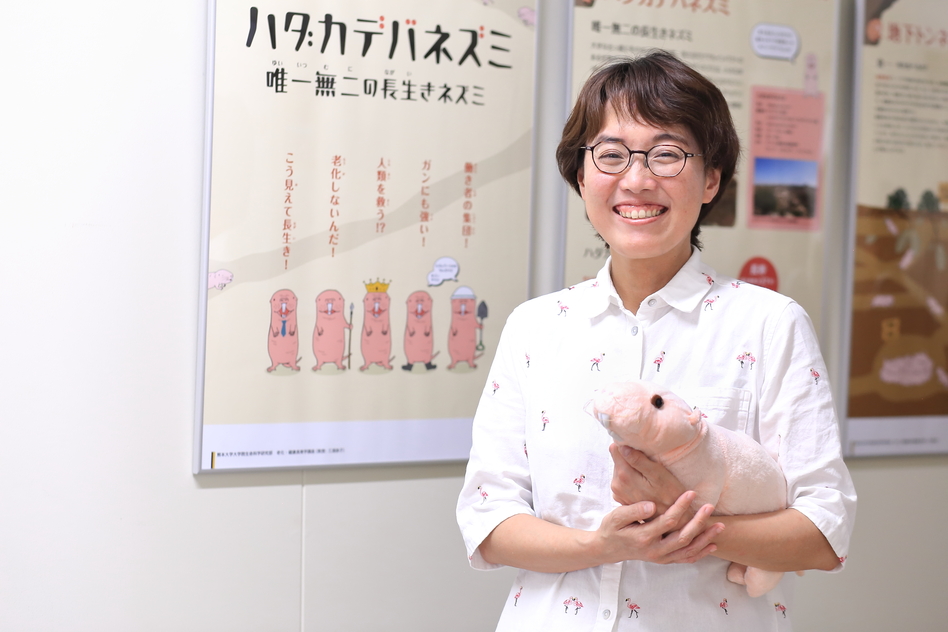
Visiting 3S Researchers #15 Dr. Kyoko MiuraDecoding the Secrets of Long-Lived, Cancer-Resistant Rats to Advance Medical Research —Pioneering the Development of Novel Model Animals—
-
 Research Grants
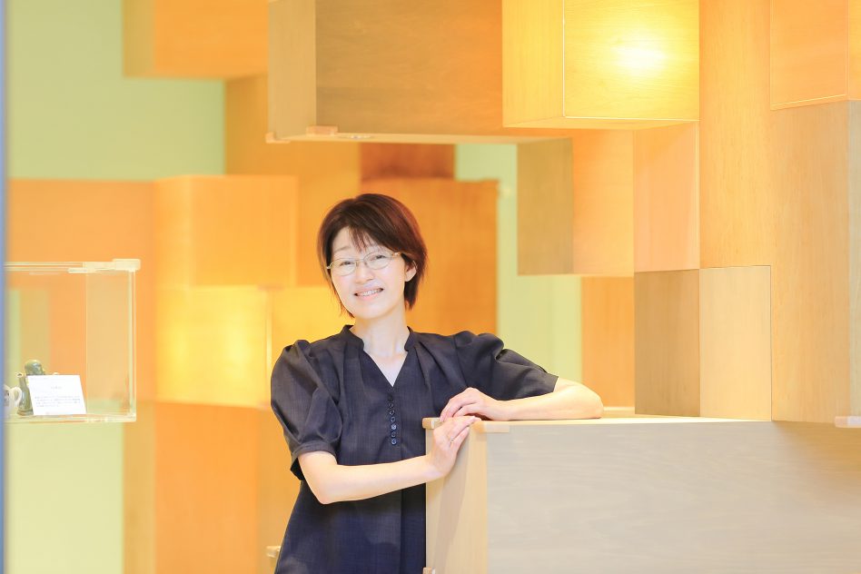
Visiting 3S Researchers #14 Dr. Megumi Sasaki Practice and Research in Safeguarding College Students’ Mental Health ─Searching for Clues to Reduce Procrastination─
-
 InformationResearch Grants
Visiting 3S Researchers #13 Dr. Kazuya SeoWhat Sports Engineering Can Do to Enhance the Appeal of Sports Events
-
 Research Grants
Visiting 3S Researchers #12 Dr. Sho TanimotoWhat Motivates Mathematicians to Tackle Difficult Problems?—Towards a Proof of Manin’s Conjecture Concerning Rational Points on Geometric Figures—